
High breast density is common. In the U.S., 40-50 percent of women ages 40-74 have dense breasts. The greater the density, the greater the risk of masking cancer on a mammogram.
Screening Breast Ultrasound
Breast Ultrasound Screening for Dense Breasts
With a specialized 3D ultrasound scan, real-time images are collected allowing radiologists to look at specific areas of dense tissue to identify breast cancers.
While a mammogram remains the primary tool for breast cancer screening, healthcare providers often order a screening ultrasound as a supplemental test to look for abnormalities that the mammogram might miss.
Other facts about breast screening with ultrasound:
- Penetrates Dense Tissue: On a mammogram, dense tissue looks white, which can completely mask cancer cells (which also appear white). Ultrasound waves pass through this tissue to reveal hidden lumps.
- Finds Missing Cancers: For every 1,000 individuals with dense breasts who have a normal mammogram, a supplemental screening ultrasound catches an additional 2 to 3 hidden cancers.
- Catches Early-Stage Invasive Cancers: More than 85% of cancers found exclusively via ultrasound are early-stage, meaning they are highly treatable before spreading.
- Zero Radiation Exposure: Unlike mammograms, ultrasounds do not use radiation. This makes them perfectly safe for repeat screenings, as well as for women who are pregnant or breastfeeding.
What is Breast Density?
While dense breast tissue is perfectly normal – affecting approximately 40 percent of the population – breast density cannot be felt and it is not related to breast size or firmness. It can only be seen on a mammogram.
Breast tissue is considered dense if there is a lot of fibrous or glandular tissue and not much fat. Fibrous and glandular tissue looks white on a mammogram, as does a possible tumor. This makes it difficult to tell the difference between a tumor and dense breast tissue on a mammogram.
Women with dense breasts should consider talking with their doctor about additional screening exams such as ABUS or Abbreviated breast MRI.
Breast Density Scale:
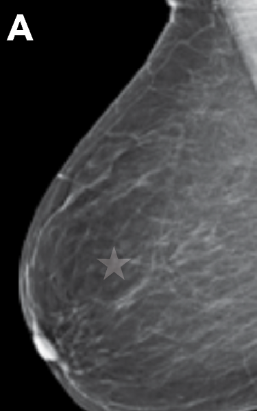
Almost entirely fatty
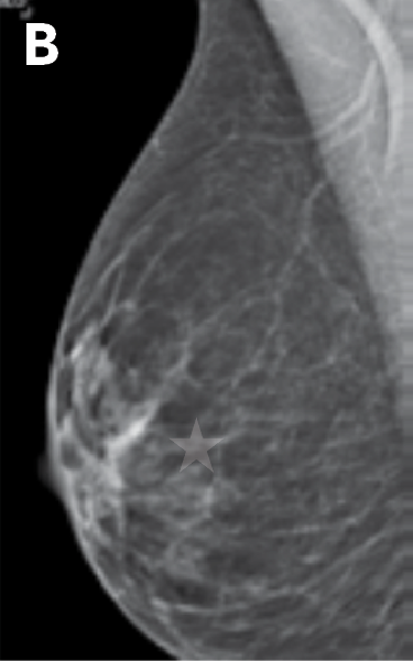
Scattered areas of fibroglandular density
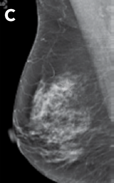
Heterogeneously dense
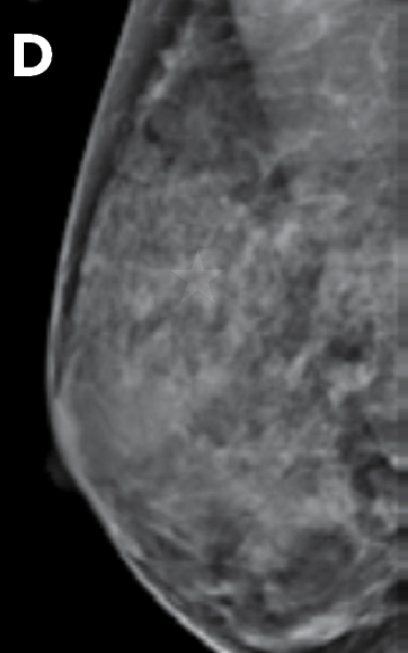
Extremely dense
Dense breasts are identified by a radiologist using mammography exams. Physical touch or self-exams cannot identify dense breasts. The mammogram offers clarity to the amount of dense tissue compared to fatty tissue and specifies what level of density the breast tissue is. The mammography report sent to your referring provider will have the details about density levels.
Radiologists classify breast density using a four-level scale (A-D). Category A is almost all fatty tissue and category B is scattered fibrous tissue. Both A and B are considered NOT DENSE. Category C is classified as heterogeneously dense and category D is extremely dense. Both C and D are considered DENSE breast tissue.
Mammography remains the gold standard for all women 40 or older. You should continue to get your mammogram annually. However, there are supplemental exam options that your doctor may recommend. Because dense tissue can hide cancers and independently increases risk, additional tests like breast ultrasound or MRI are recommended to improve detection rates. Ask your doctor if supplemental exams may be right for you. If they are, we also perform many of those exams at our outpatient center and will be happy to coordinate all your breast imaging needs.